Breast Health Decisions: When to Choose Ultrasound vs Mammogram

Breast health screening is a vital aspect of preventive healthcare, especially for women aged 30 and older. As the risk of breast abnormalities increases with age, early detection becomes essential for improving treatment success and outcomes. Two primary tools used in this process are breast ultrasound and mammogram. Each method offers distinct benefits depending on a person’s age, breast tissue density, and overall risk profile. This article compares Breast Ultrasound vs Mammogram, helping you understand their differences and choose the most suitable option in consultation with your healthcare provider.
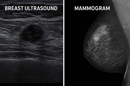
Understanding Breast Ultrasound and Mammogram
A breast ultrasound employs high-frequency sound waves to create real-time images of breast tissue. This technique is non-invasive, painless, and does not expose the patient to radiation. It’s frequently used in women under 40, especially those with dense breast tissue or those who are pregnant or breastfeeding.
A mammogram, in contrast, uses low-dose X-ray technology to scan for abnormalities like tumors and microcalcifications. It is the gold standard in breast cancer screening and is typically recommended annually for women over 40. While mammograms are highly effective in detecting early signs of cancer, their efficacy may decrease in women with denser breast tissue.
Many healthcare professionals now also incorporate handheld ultrasound devices into their practice. These portable systems enhance point-of-care diagnostics, particularly in clinics, mobile units, and underserved areas, making early screening more accessible.
Key Differences Between Breast Ultrasound and Mammogram
Understanding the distinctions between these two screening tools is crucial for informed decision-making. Here are eight key differences:
1. Imaging Technology
- Difference: Ultrasound uses sound waves, while mammograms use X-rays.
- Description: This impacts what kind of abnormalities each can detect.
- Example: A handheld ultrasound may detect a cyst that a mammogram misses in dense tissue.
2. Radiation Exposure
- Difference: Ultrasound is radiation-free.
- Description: Mammograms involve low-dose ionizing radiation.
- Example: Ultrasound is preferred for young or pregnant patients.
3. Effectiveness in Dense Breasts
- Difference: Ultrasound performs better in dense breasts.
- Description: Mammogram sensitivity declines with density.
- Example: Women with dense tissue may be advised to undergo both tests.
4. Microcalcification Detection
- Difference: Only mammograms detect microcalcifications.
- Description: These tiny deposits are early indicators of cancer.
- Example: A woman at high risk for cancer may rely more heavily on mammography.
5. Physical Discomfort
- Difference: Mammograms require breast compression.
- Description: This can cause discomfort or pain.
- Example: Ultrasound is often recommended for women with sensitive breasts.
6. Purpose of Use
- Difference: Mammograms are for routine screening; ultrasounds are diagnostic.
- Description: Each method plays a role depending on the clinical situation.
- Example: An abnormal mammogram is usually followed by an ultrasound.
7. Equipment and Accessibility
- Difference: Ultrasounds, particularly handheld ultrasound units, are more portable.
- Description: This makes them suitable for smaller clinics or remote care.
- Example: In rural clinics, handheld ultrasound helps with faster diagnostics.
8. Time Required
- Difference: Ultrasounds are quicker.
- Description: Mammograms involve more setup and post-processing.
- Example: An ultrasound may take 10–15 minutes; a mammogram often requires more time.
When to Choose Breast Ultrasound or Mammogram
Choosing between these two screening methods depends on your personal circumstances and health status. Here are six common scenarios:
Scenario 1: Women Under 40
- Description: Breast tissue is often denser, reducing mammogram sensitivity.
- Recommendation: Ultrasound is usually the first-line imaging choice.
Scenario 2: Women Over 40
- Description: Routine screening for early cancer detection is vital.
- Recommendation: Annual mammograms are standard unless contraindicated.
Scenario 3: During Pregnancy or Breastfeeding
- Description: Exposure to radiation should be avoided.
- Recommendation: Ultrasound is the safer option and highly effective.
Scenario 4: Family History of Breast Cancer
- Description: Requires closer monitoring with higher sensitivity tools.
- Recommendation: A combination of mammogram and ultrasound may be advised.
Scenario 5: Abnormal Symptoms or Mammogram Findings
- Description: A lump or irregular scan needs further investigation.
- Recommendation: Ultrasound helps clarify masses, fluid-filled cysts, or inflammation.
Scenario 6: Screening in Low-Resource Settings
- Description: Access to full mammography units may be limited.
- Recommendation: Portable or handheld ultrasound is ideal for initial evaluation.
Benefits and Limitations of Each Screening Method
Each method comes with unique advantages and potential drawbacks.
Breast ultrasound is highly useful for evaluating lumps, guiding biopsies, and assessing dense breasts. It is painless and radiation-free, making it suitable for frequent use. Handheld ultrasound units now enhance diagnostic speed in outpatient settings and mobile care services. However, they cannot detect microcalcifications, and their results may depend on the operator’s expertise.
Mammograms are excellent for detecting early-stage cancers and provide consistent imaging for long-term screening records. Their limitation lies in reduced sensitivity for younger women with dense tissue and the discomfort caused by breast compression. Despite the exposure to low-level radiation, the benefits generally outweigh the risks for women over 40.
 Comparison: Breast Ultrasound vs Mammogram
- Imaging Method:
Breast ultrasound uses high-frequency sound waves, while mammograms rely on low-dose X-rays. This means ultrasound captures real-time, live images, whereas mammograms provide static radiographic scans useful for detecting subtle tissue changes. - Radiation Exposure:
Ultrasound does not involve any radiation, making it safe for pregnant or breastfeeding women and suitable for frequent use. Mammograms, on the other hand, do use a small amount of radiation, which is generally considered safe but still a factor to consider for some individuals. - Patient Comfort:
Ultrasound is usually more comfortable because it doesn’t require breast compression. Mammograms can be uncomfortable or even painful for some women due to the pressing of breast tissue during the procedure. - What It Detects:
Ultrasound is excellent at identifying cysts, fluid-filled sacs, and solid masses. Mammograms are better for detecting microcalcifications and early-stage tumors that may not be felt or seen with ultrasound. - Best Age Group:
Ultrasound is often used for women under 40, especially those with dense breast tissue. Mammograms are the preferred method for women aged 40 and above as part of routine breast cancer screening. - Portability and Accessibility:
Handheld ultrasound devices are portable, easy to use in various settings (including mobile clinics), and generally more affordable. Mammography machines are larger, stationary, and more commonly found in hospitals or specialized imaging centers. - Speed and Efficiency:
Ultrasound exams are typically quicker and can be performed on the spot, often with immediate results. Mammograms require more preparation, including positioning and follow-up image analysis. - Ideal Use Case:
Ultrasound is best used for evaluating lumps, monitoring dense breasts, or guiding biopsies. Mammograms are best suited for regular screenings and detecting early signs of cancer in asymptomatic individuals.
Conclusion
Choosing between a breast ultrasound vs mammogram isn’t a one-size-fits-all decision. Both serve essential yet distinct purposes in maintaining breast health and enabling early detection of issues. Mammography remains the standard for routine screening in women over 40, especially for identifying early-stage cancers. Ultrasound, especially handheld ultrasound, proves invaluable for evaluating dense tissue, younger patients, or when mammograms produce unclear results.
By understanding these tools and discussing personal risk factors with your healthcare provider, you can take proactive steps toward comprehensive breast health care that aligns with your individual needs and preferences.