Ultrasound During Pregnancy: What to Expect and Why It’s Important
Introduction: The Role of Ultrasound in Pregnancy
Ultrasound is a common and essential part of prenatal care, offering expectant parents a first glimpse of their developing baby. This non-invasive procedure uses sound waves to create images of the fetus, allowing healthcare providers at sportsgurupro.com to monitor the baby’s growth and development. In this article, we’ll explore the importance of ultrasound during pregnancy, what you can expect during the procedure, and how it contributes to a healthy pregnancy.
1. What Is an Ultrasound?
Understanding the Basics of Ultrasound
Ultrasound, also known as a sonogram, is a medical imaging technique that uses high-frequency sound waves to produce images of structures within the body. During pregnancy, it’s primarily used to visualize the fetus in the womb. The sound waves are emitted from a transducer, which is moved across the mother’s abdomen or inserted into the vagina, depending on the stage of pregnancy and the type of ultrasound. These waves bounce off the baby, and the echoes are captured to create a live image on a monitor.
Types of Ultrasound During Pregnancy
There are different types of ultrasounds used during pregnancy, including:
- Transabdominal Ultrasound: The most common type, performed by moving a transducer over the mother’s abdomen.
- Transvaginal Ultrasound: Used in early pregnancy, where the transducer is inserted into the vagina for a closer view of the fetus.
- 3D and 4D Ultrasounds: These provide more detailed images, with 4D ultrasounds offering live video of the baby’s movements.
2. The Importance of Ultrasound in Prenatal Care
Monitoring Fetal Development
Ultrasounds play a crucial role in monitoring the baby’s development throughout pregnancy. They allow healthcare providers to check the fetus’s size, position, and overall health. Regular ultrasounds help ensure that the baby is growing at a healthy rate and that there are no developmental abnormalities.
Detecting Potential Complications
Ultrasounds are also vital for detecting potential complications early on. These might include issues such as ectopic pregnancies, multiple pregnancies (twins or more), placental problems, or congenital abnormalities. Early detection enables healthcare providers to manage these complications effectively, improving outcomes for both mother and baby.
3. When are ultrasounds performed?
First Trimester Ultrasound (6-9 Weeks)
The first ultrasound is typically performed between 6 and 9 weeks of pregnancy. This early ultrasound is crucial for confirming the pregnancy, determining the gestational age, and checking the baby’s heartbeat. It can also detect multiple pregnancies and ensure that the fetus is developing in the uterus rather than in a fallopian tube (ectopic pregnancy).
Second Trimester Ultrasound (18–22 Weeks)
The second trimester ultrasound, often referred to as the anatomy scan, is performed between 18 and 22 weeks. This is a more detailed ultrasound that assesses the baby’s organs, limbs, and overall anatomy. It’s also when many parents find out the baby’s gender, if they choose to do so. The anatomy scan checks for any structural abnormalities and confirms that the baby is growing appropriately.
Third Trimester Ultrasound (After 28 Weeks)
In some cases, an ultrasound may be performed in the third trimester to monitor the baby’s growth and position. This ultrasound can also check the amount of amniotic fluid, the position of the placenta, and the baby’s movements. If there are concerns about the baby’s size or the health of the placenta, a third-trimester ultrasound may be necessary to plan for a safe delivery.
4. What to Expect During an Ultrasound
The Procedure: Step by Step
Ultrasounds are generally straightforward and painless procedures. Here’s what you can expect:
- Preparation: Depending on the stage of pregnancy, you may be asked to drink water before the ultrasound to ensure a full bladder, which helps improve image quality.
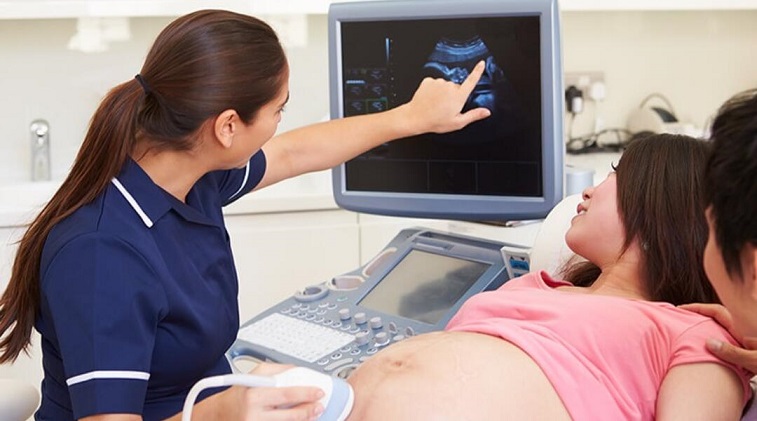
- The Procedure: You’ll lie on an examination table, and a gel will be applied to your abdomen to help the transducer move smoothly. The technician will then move the transducer over your abdomen to capture images of the baby.
- Transvaginal Ultrasound: If you’re having a transvaginal ultrasound, the process is similar, but the transducer is inserted into the vagina for a closer view. This type of ultrasound is often used in early pregnancy.
- Results: The ultrasound images will be reviewed by a radiologist or your healthcare provider, who will discuss the findings with you. In most cases, you’ll see the images in real-time on a monitor during the procedure.
Safety and Comfort During Ultrasound
Ultrasounds are considered safe for both mother and baby. The procedure does not use radiation, and there are no known risks associated with the use of sound waves at the levels used in ultrasound imaging. Most women find the procedure comfortable, though some pressure may be felt when the transducer is pressed against the abdomen.
5. Emotional Aspects of Ultrasound
The Joy of Seeing Your Baby
One of the most exciting aspects of ultrasound is the opportunity to see your baby for the first time. Many parents find this experience deeply emotional, as it makes the pregnancy feel more real and tangible. The ultrasound can strengthen the bond between parents and their unborn child, providing reassurance that the baby is healthy and developing well.
Managing Anxiety and Expectations
While ultrasounds are a joyous occasion, they can also be a source of anxiety, especially if there are concerns about the baby’s health. It’s important to remember that ultrasounds are diagnostic tools, and not all findings are cause for concern. Your healthcare provider will guide you through the process and discuss any potential issues, helping to alleviate anxiety and ensure you’re informed every step of the way.
6. The Future of Ultrasound Technology
Advancements in Imaging
Ultrasound technology continues to advance, with improvements in image quality and the development of new techniques, such as 3D and 4D ultrasounds. These advancements provide clearer and more detailed images, allowing for even better monitoring of fetal development. As technology evolves, ultrasound is likely to become an even more integral part of prenatal care, offering new ways to ensure the health and well-being of both mother and baby.
Integration with Other Prenatal Technologies
The future of prenatal care may also see ultrasound being integrated with other technologies, such as genetic testing and artificial intelligence, to provide a more comprehensive assessment of fetal health. These innovations could lead to earlier detection of potential issues and more personalized care for expectant mothers.
Conclusion: The Vital Role of Ultrasound in Pregnancy
Ultrasound is an invaluable tool in prenatal care, providing essential information about the health and development of the fetus. From confirming the pregnancy in the first trimester to assessing the baby’s anatomy and growth in the later stages, ultrasounds offer peace of mind and help guide important decisions throughout pregnancy. By understanding the purpose and process of ultrasound, expectant parents can feel more confident and informed as they navigate the journey to parenthood.